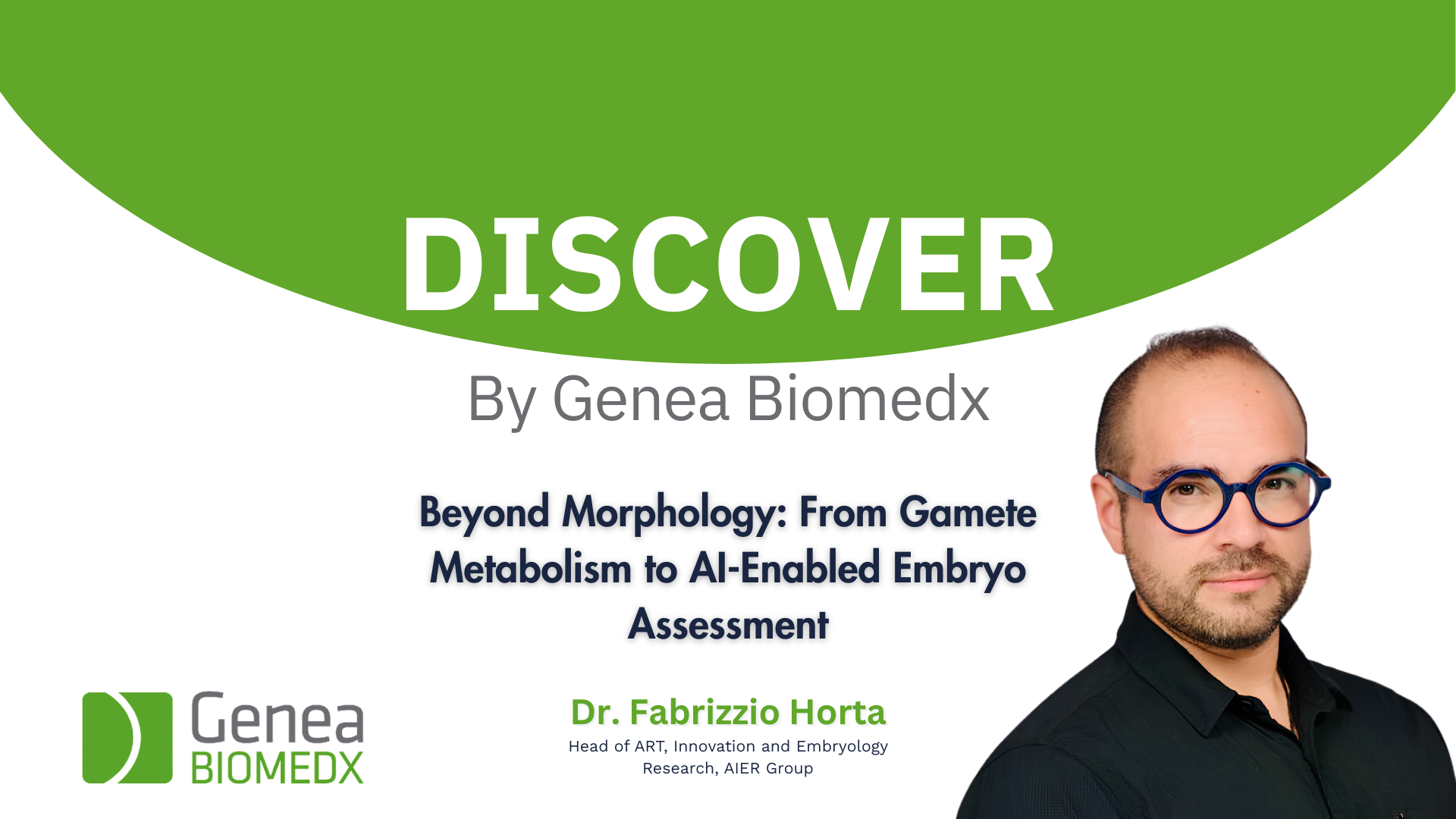
Beyond Morphology: From Gamete Metabolism to AI-Enabled Embryo Assessment
Dr. Fabrizzio Horta
Head of ART, Innovation and Embryology Research, AIER Group
Discover how label-free metabolic imaging and artificial intelligence guide embryologists to objective viability insights and standardise embryo ranking.
What you will learn:
- The AI Advantage: How deep learning converts complex metabolic imaging and time-lapse data into clinically interpretable decision-support outputs.
- Key Metabolic Tracking: The specific redox metrics, NAD(P)H/FAD signatures, and ‘quiet embryo’ principles detected by advanced label-free microscopy.
- Custom Scoring Flexibility: How to integrate metabolic readouts with lab-specific culture conditions and proprietary algorithms to personalise embryo ranking.
Synergising Metabolic Imaging for AI-Enabled Embryo Assessment
Advanced microscopy generates complex metabolic datasets that are difficult to interpret manually alongside time-lapse morphokinetic data. Moreover, integrating artificial intelligence bridges this analytical gap by automatically extracting and quantifying spatial metabolic heterogeneity.
Unlocking the ‘Quiet Embryo’ Hypothesis
Because traditional morphology cannot fully capture embryonic physiological health, manual assessment remains limited in predicting developmental viability. Consequently, label-free metabolic imaging provides a non-invasive strategy to assess cellular redox balance through endogenous autofluorescence. For example, this approach validates the ‘quiet embryo’ hypothesis, revealing that optimal viability is linked to a regulated metabolic range rather than maximal activity.
Advancing Non-Invasive Decision Support in ART
Ultimately, the rising demand for effective assisted reproductive technologies requires reliable, non-invasive alternatives to complement current selection strategies. Therefore, metabolic imaging paired with AI offers a robust decision-support tool to risk-stratify embryos and optimise transfer strategies.
Related Articles
-
Transforming Embryo Culture: Insights and Innovations with the GERI Incubator
Discover how Time-Lapse incubation is transforming embryo culture, helping IVF labs make smarter decisions, optimise workflows, and improve clinical outcomes.
-
Beyond Morphology: From Gamete Metabolism to AI-Enabled Embryo Assessment
Discover how label-free metabolic imaging and artificial intelligence guide embryologists to objective viability insights and standardise embryo ranking.
-
Trehalose-Based Vitrification
Expert insights into next-generation cryopreservation strategies for the modern IVF laboratory